Heart House News

New Medical Technology Provides High-Quality Images, Lower Radiation
By Kim Mulford
Courier-Post | August 31, 2013
Changes in health care tests are providing better information about what's going on inside the body - creating clearer images while often using lower radiation doses.
That could mean earlier detection, fewer tests, a reduction in unnecessary surgeries and increased accuracy when diagnosing women and heavier patients.
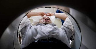 Mark Evans of Levittown, Pa., an IT administrator at The Heart House in Haddon Heights, demonstrates a PET scanner at the facility. The scan offers better views of blood flow to the heart. |
While new tests can be more expensive and are not always covered by private insurance, they have become more available to South Jersey patients, as medical practices and hospitals invest in new machines and computer software.
From 3-D mammograms to improvedscreening for lung cancer and heart disease, technology is quickly changing how doctors do their jobs.
At The Heart House in Haddon Heights, Dr. Deborah Sambucci uses the results from a new PET scan to see how cardiac patients respond to stress on the heart. The test can be used in place of a nuclear stress test, used to determine which patients need heart catheterizations.
The cardiac PET scan offers better views of blood flow to the heart, while exposing patients to less radiation than a traditional test, Sambucci said. That's important.
"As technology advances, we start radiating people more," she explained. "There's a lot more testing we can do for them. People want the tests done, and we want to do it. But it's exposing people to more and more radiation."
It takes about 45 minutes to an hour for the scan, about a third of the time required for a traditional stress test. The latter takes pictures of a patient's heart before and after exercising or taking medication to increase the heart rate. The new test takes heart images in one continuous session.
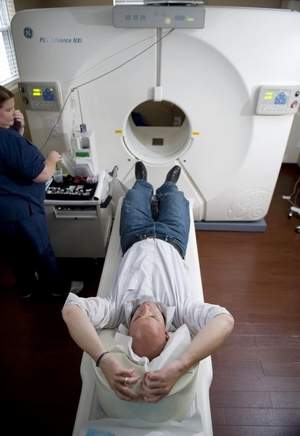 Nuclear medicine technologist Suzanne Parker of Washington Township assists Mark Evans of Levittown, PA (an IT administrator at The Heart House in Haddon Heights), as Parker demonstrates how a PET scanner is operated at The Heart House in Haddon Heights. |
The downside? It's more expensive. Medicare and some insurance companies cover it, while others are starting to come around, Sambucci said.
"We're finding more and more people are becoming comfortable with covering it."
Other advancements make it easier for patients to handle necessary testing. Larchmont Medical Imaging in Moorestown uses an extremity MRI for patients who need a scan of a limb or joint.
Instead of requiring patients to slide into a full-body scanner, the test is designed to examine just a knee, ankle, foot, hand, wrist or elbow. It's usually ordered by orthopedic doctors to see damage caused by sports injuries or arthritis.
The scanner looks something like a front-loading washer. It also creates a better picture, according to Larchmont radiologist Dr. Andrew Zeiberg.
"When you have the whole body in a closed magnet scanner, the wrist is off-center," he said. "That makes it challenging to get high-quality images.
"Extremity MRI places the wrist at the center of the bore for a clearer, more detailed view."
An investment in computer upgrades allowed Larchmont to reduce the amount of radiation needed during CT scans by 30 to 40 percent, while preserving image quality, Zeiberg said.
More complex algorithms create better images by sorting useful data from what Zeiberg calls "noise."
"It was a big investment for us, but it was purely for patient safety.''
And now that imaging studies are done digitally instead of using film, doctors can have nearly instant access to the results, instead of waiting a week or longer a decade ago.
"The flow of information is now lightning fast," Zeiberg noted.
In January, Virtua partnered with Children's Hospital of Philadelphia to open an outpatient imaging center in Voorhees. Children who need sedation during imaging can get their tests done there instead of traveling to the city.
By October, Virtua will offer a low-dose CT scan to screen for lung cancer in patients who have a history of smoking. The new test will enable pulmonologists to diagnose patients at earlier stages of the disease, before symptoms appear.
Typically, a chest X-ray uncovers lung cancer when it is in the middle or late stages, said Barry Graf, vice president of integrated operations for Virtua. Studies have shown earlier detection leads to "much greater survival rates" for lung cancer patients.
Better breast imaging is now available, too. South Jersey Radiology in Voorhees offers 3-D mammograms, also called digital breast tomosynthesis.
The test uses slightly more radiation than a traditional mammogram, but offers clearer pictures of suspicious tissue. Work is underway to create such images using less radiation.
Cooper Cancer Institute uses contrast enhanced spectral mammography to see unusual blood flow patterns within the breast to find cancer. The five-minute test is used after an inconclusive mammogram or ultrasound.
Cooper started using the technology last November, and was among the first sites nationally to offer it.
But while more imaging tools are available to doctors and their patients, it's important to choose the appropriate tests, Graf said.
"The pressures in health care today are around making a diagnosis as quickly and safely as possible."
[To read this article online, follow this link.]